CÁNDIDA SPP

Gallery
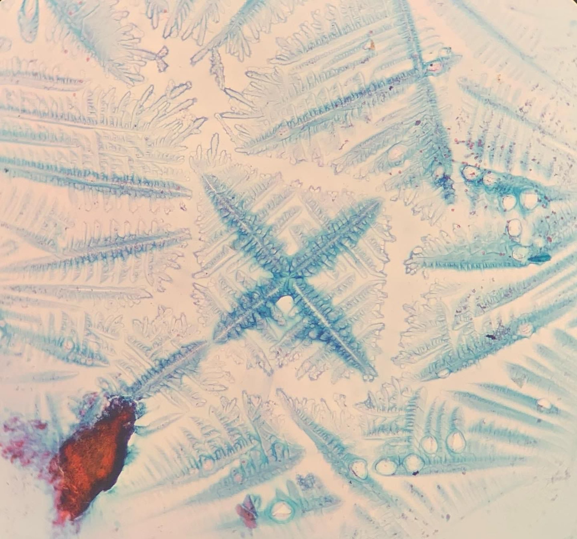
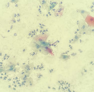
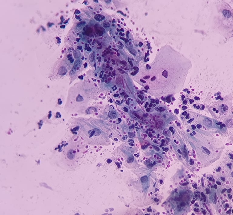
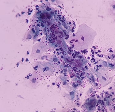
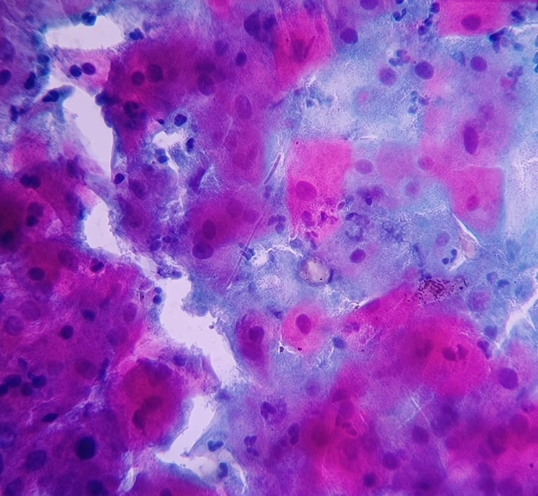
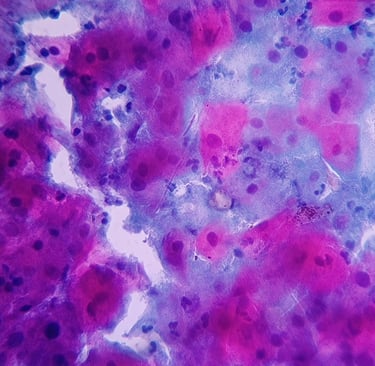
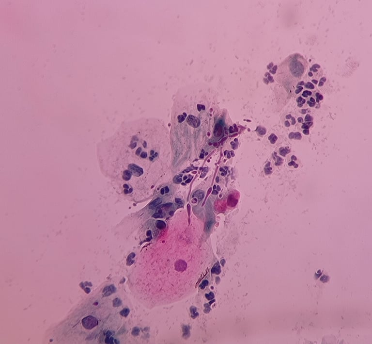
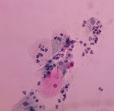
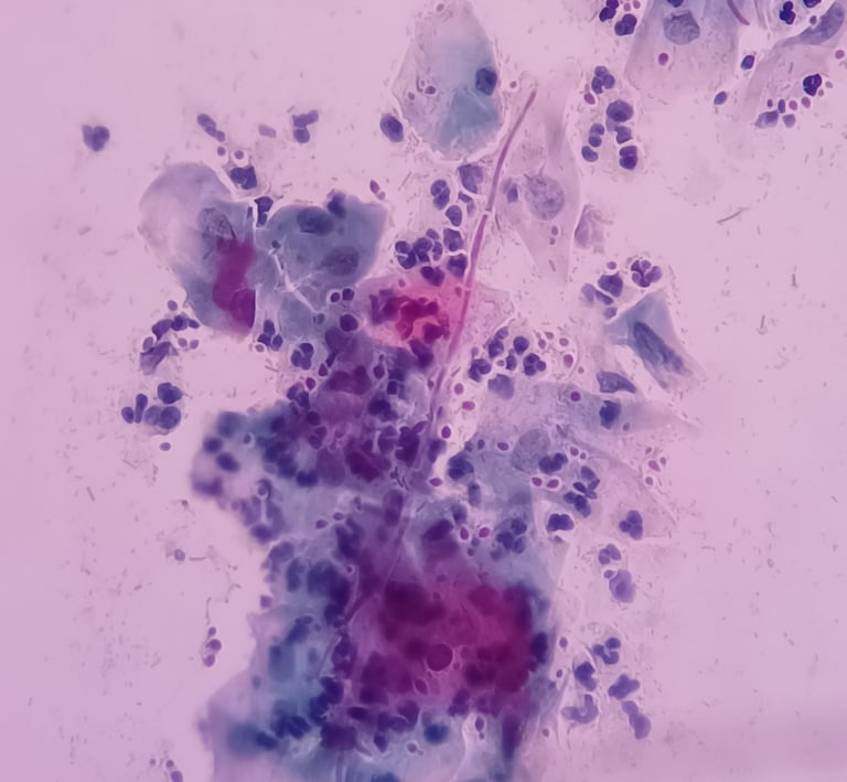
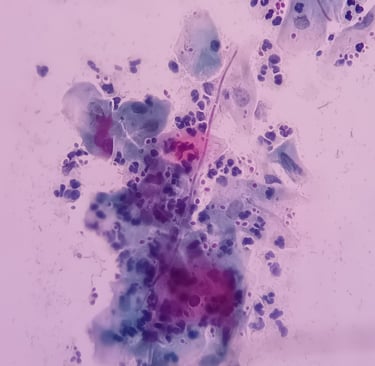
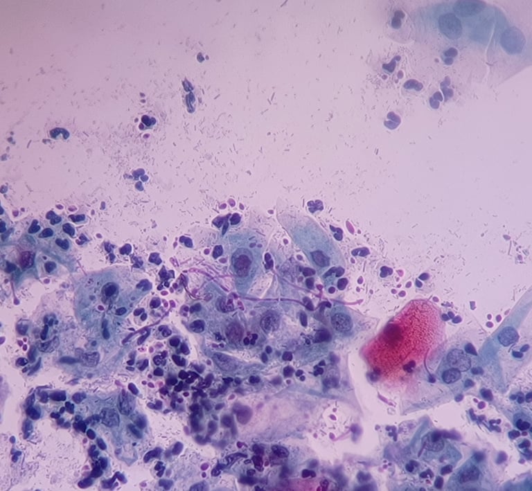
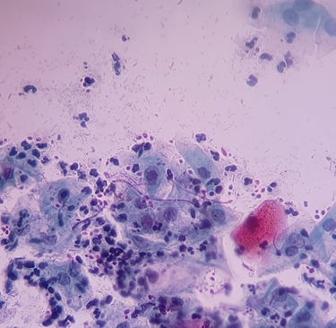
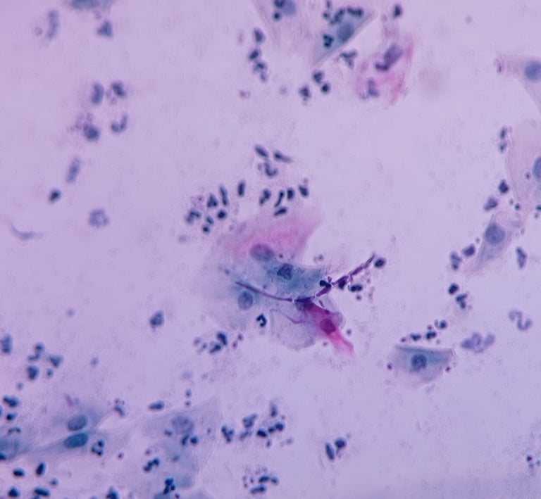
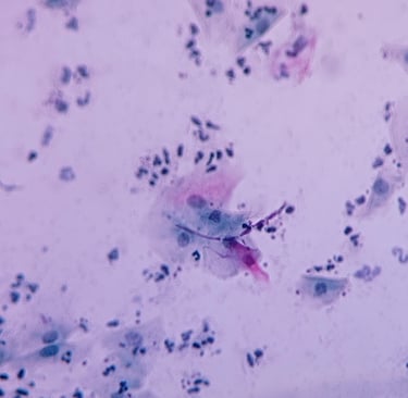
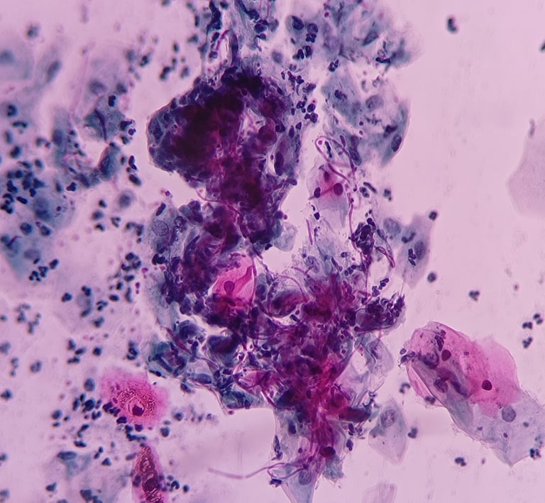
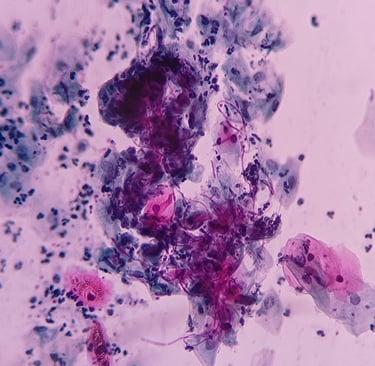
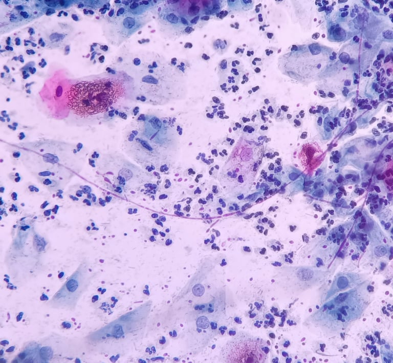
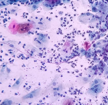
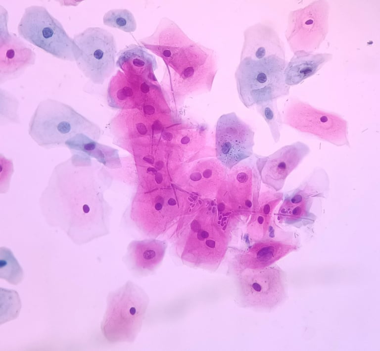
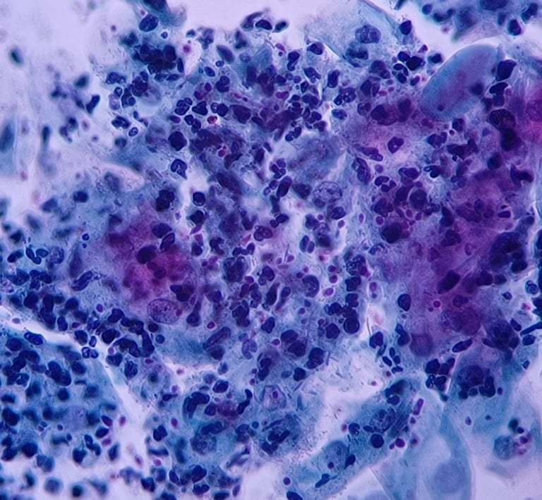
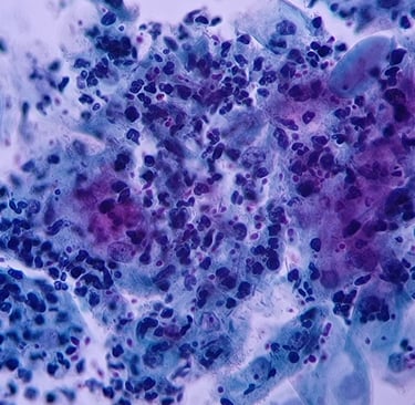
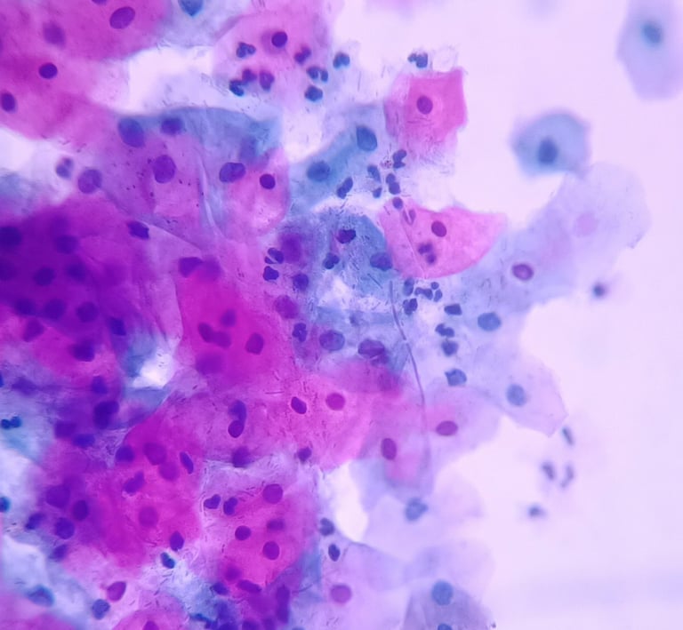
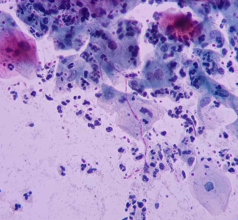
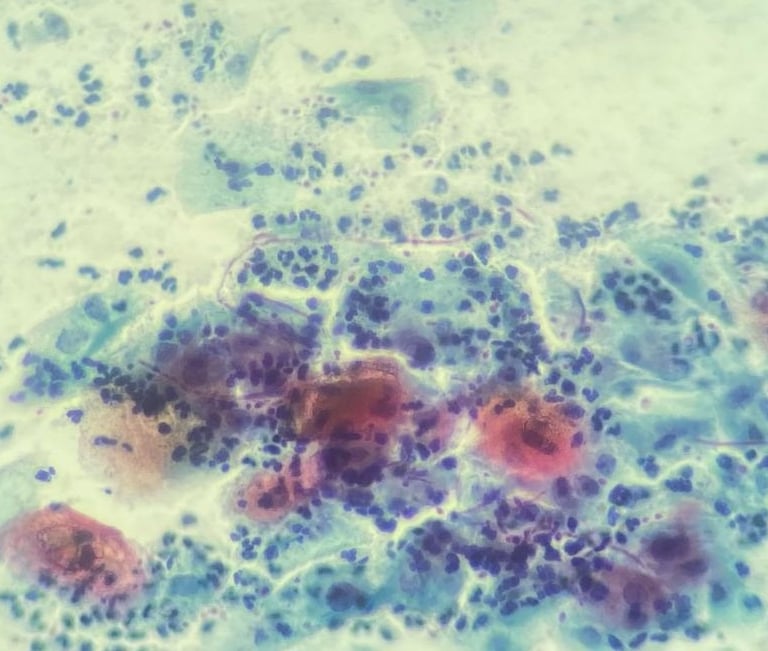
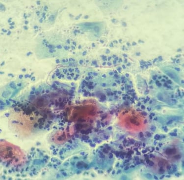
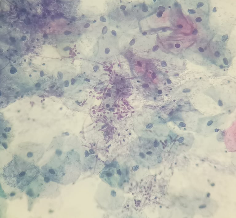
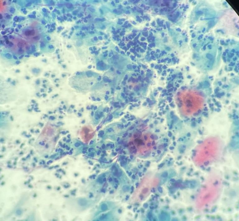
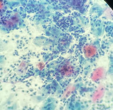
“Budding yeast (3–7 μm) and/or pseudohyphae; pseudohyphae can be quite long, spanning many cells, and are eosinophilic to gray brown on the Papanicolaou stain.
Pseudohyphae, formed by cytoplasmic extension of budding yeasts, lack true septations but show complete constrictions along their length that indicate the formation of new cells.
Fragmented leukocyte nuclei and groups of squamous epithelial cells ‘speared’ by pseudohyphae and held together in a rouleaux are often seen.”
— Nayar, R. & Wilbur, D.C. (Eds.). The Bethesda System for Reporting Cervical Cytology: Definitions, Criteria, and Explanatory Notes. 3rd ed. Springer, 2015.
This website is not affiliated with or endorsed by the authors, editors, or publishers of The Bethesda System for Reporting Cervical Cytology. Content quoted is used strictly for educational and informational purposes.


Suggestions or technical problems
Send us your suggestions for improving the site or any technical issues you may have. Thank you...